Welcome to the Human Anatomy Laboratory Manual with Cat Dissections, a comprehensive guide designed to enhance understanding of human anatomy through hands-on feline dissection․ This manual provides a structured approach to exploring anatomical structures, aligning with course objectives and offering practical experience for students․ By combining detailed instructions with visual aids, it ensures a deeper appreciation of biological systems and their functions․
1․1 Importance of Cat Dissections in Anatomy Education
Cat dissections are highly valued in anatomy education for their similarity to human anatomy, providing students with hands-on experience․ They bridge theoretical knowledge with practical skills, enhancing understanding of complex anatomical structures and their functions․ This approach fosters critical thinking and prepares learners for real-world applications in medical and scientific fields effectively․
1․2 Historical Context of Cat Dissections in Laboratory Settings
Cat dissections have been a cornerstone of anatomy education for centuries, dating back to early medical training․ Their anatomical similarity to humans and accessibility made them ideal for laboratory use․ This tradition has evolved, with modern manuals refining techniques and aligning with educational standards to enhance student learning experiences․
1․3 Aligning the Manual with Course Objectives
This manual is carefully designed to align with course objectives, ensuring a comprehensive understanding of human anatomy․ It integrates practical dissection exercises with theoretical knowledge, covering all major body systems․ Each activity is tailored to reinforce key concepts, providing students with hands-on experience and preparing them for clinical applications in anatomy․
Preparation for Cat Dissection
Proper preparation is essential for a successful cat dissection․ This includes gathering tools, following safety protocols, and understanding ethical considerations to ensure a respectful and effective learning experience․
2․1 Safety Protocols and Laboratory Etiquette
Adhering to safety protocols is crucial in cat dissection․ Wear gloves, goggles, and lab coats to prevent exposure․ Handle tools carefully and ensure proper clean-up․ Follow biohazard disposal guidelines and maintain a respectful, organized workspace․ Understanding laboratory etiquette promotes a safe and efficient learning environment for all participants․
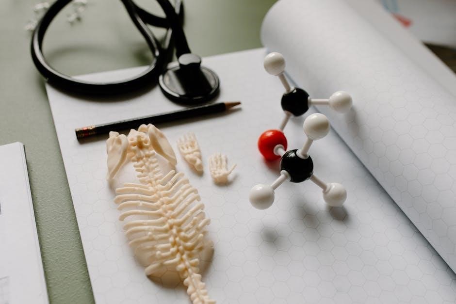
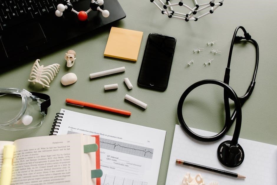
2․2 Essential Tools and Equipment for Dissection
The dissection process requires specific tools, including scalpels, forceps, scissors, and dissecting trays․ Additional equipment like probes and magnifying glasses aid in detailed examination․ Proper use and maintenance of these tools ensure precision and safety during laboratory exercises, facilitating a successful and precise dissection experience․
2․3 Ethical Considerations and Specimen Preparation
Ethical considerations in dissection emphasize respect for specimens and adherence to institutional guidelines․ Proper preparation involves handling and storing specimens safely, ensuring hygiene, and minimizing waste; This approach balances educational needs with ethical responsibility, fostering a respectful and sustainable learning environment for anatomy students․

Exploring the Skin and Skeletal System
This chapter delves into the structure and function of the skin and skeletal system in cats․ Through detailed dissection, students explore skin layers, skeletal features, and their interconnections, gaining practical insights into their roles in movement and protection․
3․1 Skin Structure and Its Functions
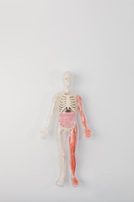
The skin is the body’s largest organ, serving as a protective barrier․ In cats, it consists of the epidermis, dermis, and hypodermis․ This chapter examines the histological layers, sensory receptors, and specialized features like fur and paw pads, highlighting their roles in protection, thermoregulation, and sensory perception․
- Epidermis: Outermost layer, providing a barrier against pathogens․
- Dermis: Contains blood vessels, nerve endings, and hair follicles․
- Hypodermis: Connects skin to underlying tissues, aiding in movement․
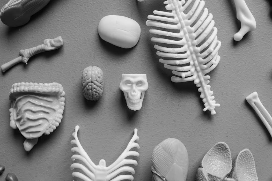
3․2 Identifying Skeletal Features in Cats
Cat skeletons consist of the axial and appendicular systems, adapted for flexibility and agility․ Key features include the skull, vertebral column, ribcage, and sternum․ The appendicular skeleton includes the upper and lower limbs․ Notable bones are the hyoid apparatus, zygomatic arches, and mandible․ The radius and ulna fuse in the forearm, while the tibia and fibula support the lower leg, enabling movement and weight-bearing functions․
3․3 Dissection Techniques for Skin and Skeleton
Begin with precise skin incisions using a scalpel, carefully retracting tissue with forceps to avoid damaging underlying structures․ For skeletal access, employ bone saws or osteotomes․ Work methodically, ensuring clear exposure of bones and joints․ Maintain a well-lit workspace and use magnification to observe fine details, enhancing your understanding of feline anatomy․
Muscular System Dissection
Begin with chest dissection, using a scalpel and forceps to expose muscles․ Retract tissue carefully, employing magnification to observe muscle structure and attachments, aiding in understanding human anatomy through feline dissection․
4․1 Overview of Cat Musculature
Cat musculature offers insights into human anatomy, with skeletal muscles arranged into flexors and extensors․ This organization mirrors human anatomy, making feline dissection an effective teaching tool․ The muscles are grouped to facilitate movement, with distinct layers observable during dissection, aiding in the study of their structure, attachments, and functional roles․
4․2 Identifying Major Muscle Groups
The primary muscle groups in cats include flexors and extensors, which control movement․ Key muscles like the deltoid, biceps brachii, and quadriceps are easily identifiable․ These groups mirror human musculature, making feline dissection an excellent model for studying muscle structure, attachments, and functional roles in locomotion and posture․
4․3 Muscle Function and Movement Analysis
Muscles are essential for movement, with flexors and extensors enabling joint mobility․ In cats, these muscles facilitate walking, running, and climbing․ By analyzing muscle attachments and contractions, students can understand how skeletal muscles contribute to motion․ This practical approach helps correlate feline anatomy with human musculature, enhancing comprehension of muscle function and biomechanics․

Nervous System Exploration
Exploring the nervous system through cat dissection reveals its intricate structure, including the brain, spinal cord, nerves, and ganglia․ This hands-on approach aids in understanding human neural functions and anatomical correlations, essential for veterinary and medical applications․
5․1 Structure of the Cat Nervous System
The cat nervous system mirrors human anatomy, consisting of the central and peripheral nervous systems․ Dissection enables detailed examination of the brain, spinal cord, nerves, and ganglia, offering insights into neural pathways and anatomical features crucial for veterinary medicine and human health studies․
5․2 Identifying Key Nerves and Ganglia
During dissection, students identify key nerves such as the sciatic nerve and sympathetic chain, and locate cervical ganglia․ These structures are vital for understanding neural pathways and motor functions․ Proper dissection techniques ensure accurate identification, aiding in the comprehension of the peripheral nervous system’s role in both feline and human anatomy․
5;3 Dissection of the Brain and Spinal Cord
The dissection of the brain and spinal cord requires careful removal of the skull and dorsal vertebrae․ Using a scalpel and forceps, students expose these structures, preserving cranial nerves and spinal cord integrity․ This step provides insights into the central nervous system’s organization, enabling comparisons between feline and human neuroanatomy for educational clarity․
Circulatory and Respiratory Systems
This chapter explores the cat’s circulatory and respiratory systems, focusing on the thoracic cavity, heart, blood vessels, and lungs․ It examines how these structures function together․
6․1 Heart and Blood Vessel Anatomy
The feline heart consists of four chambers: two atria and two ventricles, with valves ensuring unidirectional blood flow․ Blood vessels, including arteries, veins, and capillaries, transport oxygenated and deoxygenated blood throughout the body․ This section details the structural and functional anatomy of these circulatory components, aiding in understanding their roles in maintaining life․
6․2 Lung Structure and Respiratory Mechanisms
The feline lungs are divided into lobes, each enclosed by pleura, facilitating gas exchange in the alveoli․ Respiratory mechanisms involve the diaphragm and intercostal muscles, enabling inhalation and exhalation․ This section explores the structural and functional aspects of the lungs, highlighting their critical role in oxygenation and carbon dioxide removal․
6․3 Dissection of the Thoracic Cavity
Dissection of the thoracic cavity involves carefully removing the sternum and ribs to expose the heart, lungs, and major blood vessels․ Students observe the diaphragm and its attachment to the ribcage, which plays a crucial role in respiration․ This step-by-step process highlights the structural relationships within the chest cavity․
Key landmarks include the trachea, esophagus, and aorta․ Proper use of scalpels and forceps ensures safe and precise exploration, allowing students to identify and understand the functional anatomy of the thoracic organs and their interconnections․

Digestive and Urinary Systems
This chapter explores the anatomy of the digestive and urinary systems through cat dissection․ Students examine the gastrointestinal tract, liver, pancreas, and urinary organs, gaining insights into their structure and function․ Hands-on dissection provides a comparative understanding of these systems in cats and their relevance to human anatomy․
7․1 Anatomy of the Gastrointestinal Tract
The gastrointestinal (GI) tract in cats, like in humans, consists of the mouth, esophagus, stomach, small intestine, and large intestine․ Dissection reveals the mucosa, submucosa, muscularis, and serosa layers․ The stomach’s gastric glands and the intestines’ villi are key structures for digestion and nutrient absorption, showcasing functional anatomy through comparative dissection․
7․2 Structure and Function of the Liver and Pancreas
The liver, divided into lobes, performs detoxification, metabolism, and bile production․ The pancreas consists of exocrine cells producing digestive enzymes and endocrine cells releasing hormones like insulin․ Dissection highlights their roles in digestion and metabolism, offering insights into their shared functions in both feline and human anatomy for comparative study․
7․3 Exploring the Urinary System in Cats
The urinary system in cats includes kidneys, ureters, bladder, and urethra․ Dissection reveals how kidneys filter blood to produce urine, which is stored in the bladder and expelled through the urethra․ This process mirrors human anatomy, emphasizing shared physiological mechanisms in waste elimination and fluid balance․
Reproductive System Dissection
This section guides the exploration of the feline reproductive system, examining both male and female anatomy to provide insights into mammalian reproduction and its human anatomical correlations․
8․1 Male and Female Reproductive Organs
This section details the anatomy of feline reproductive systems, focusing on the male and female organs․ Males include the testes, epididymis, and penis, while females comprise the ovaries, uterus, and vagina․ Dissection highlights their structure, function, and role in reproduction, offering insights into mammalian reproductive biology and its correlation to human anatomy․
8․2 Identifying Reproductive Structures
During dissection, identify key reproductive structures such as the testes, ovaries, uterus, and vagina․ Note the location and morphology of each organ․ Use dissection tools to carefully expose and examine these structures, ensuring accurate identification and understanding of their roles in feline reproduction․
8․3 Dissection Techniques for Reproductive Systems
Use precision instruments to carefully expose reproductive organs, making strategic incisions to avoid damaging surrounding tissues․ For males, locate the testes and epididymis, while for females, identify the ovaries, uterus, and vagina․ Employ magnification tools to observe intricate structures and ensure accurate dissection and identification of reproductive anatomy․
Microscopy and Histology
Master microscopic techniques to examine tissue samples, identifying cellular structures and correlating findings with dissection observations․ Use Boolean operators and truncation in searches to locate relevant histological resources efficiently․
9․1 Preparing Tissue Samples for Microscopy
Learn to prepare tissue samples for microscopy by fixing, sectioning, and staining․ Proper techniques ensure clear visualization of cellular structures․ Use search strategies with Boolean operators to find detailed protocols and resources for effective sample preparation in feline anatomy studies․
9․2 Identifying Cellular Structures
Master the identification of cellular structures under a microscope, focusing on key components like cell membranes, nuclei, and organelles․ Use search techniques to find resources on cellular anatomy․ Correlate microscopic observations with dissection findings to deepen your understanding of tissue organization and function in feline anatomy studies․
9․3 Correlating Dissection Findings with Microscopic Observations
This section emphasizes linking macroscopic dissection observations with microscopic cellular details․ By examining tissue samples under a microscope, students can identify structural features that align with anatomical discoveries from cat dissections․ This integration enhances understanding of how cellular structures contribute to organ function and overall feline anatomy․
Clinical Correlations and Applications
This section bridges anatomical knowledge with practical applications in veterinary medicine, using feline dissections to illustrate clinical case studies and current research, enhancing real-world understanding and skills․
10․1 Applying Anatomical Knowledge to Veterinary Medicine
This section explores how anatomical insights from cat dissections translate into practical veterinary applications, aiding in diagnosis, treatment, and surgical procedures․ By correlating feline anatomy with clinical scenarios, students gain essential skills for real-world veterinary care, bridging the gap between laboratory learning and professional practice․
10․2 Case Studies in Feline Anatomy
This section presents real-world case studies that illustrate the practical application of feline anatomy․ Through detailed examinations of specific anatomical conditions and surgical procedures, students gain hands-on experience in identifying and understanding structural abnormalities, enhancing their diagnostic and analytical skills in a clinical context․
10․3 Current Research in Cat Anatomy and Its Implications
Recent studies in feline anatomy highlight advancements in understanding regenerative processes, musculoskeletal adaptations, and neural systems․ These findings offer insights into human anatomical research, particularly in joint diseases and nervous system disorders․ Such research bridges veterinary and human medicine, fostering interdisciplinary applications and innovative treatments across species․
Safety and Disposal Protocols
Adhering to safety protocols is crucial in anatomy labs․ Proper handling of tools, biohazard disposal, and cleaning ensure a safe environment․ Always follow laboratory guidelines strictly․
11․1 Proper Handling of Dissection Tools
Always handle dissection tools with care to ensure safety and longevity․ Use gloves, store instruments in designated cases, and clean them regularly․ Scalpels and forceps require special attention to maintain sharpness and precision․ Proper handling prevents accidents and extends tool life, ensuring efficient lab operations and student safety․
11․2 Biohazard Disposal and Clean-Up
Properly dispose of biohazardous waste in sealed containers and clean surfaces with disinfectants․ Wear gloves and follow safety protocols to prevent contamination․ Ensure all materials are disinfected, and dispose of specimens according to lab guidelines․ This maintains a safe environment and adheres to regulatory standards for handling biological materials responsibly․
11․3 Laboratory Safety Best Practices
Adhere to safety protocols by wearing protective gear like gloves and goggles․ Handle sharp tools carefully and store chemicals properly․ Keep emergency equipment accessible and maintain a clean workspace․ Follow proper procedures for spills and disposal to ensure a safe learning environment for all participants․
This manual provides a hands-on approach to understanding human anatomy through cat dissections․ For further study, explore recommended textbooks, online databases, and anatomical resources for deeper insights․
12․1 Summarizing Key Concepts
This manual provides a comprehensive overview of human anatomy through cat dissection, covering skin, skeletal, muscular, nervous, circulatory, respiratory, digestive, urinary, and reproductive systems․ It emphasizes hands-on techniques, essential tools, and ethical considerations, aligning with course objectives to foster practical understanding and application of anatomical knowledge․
12․3 Encouraging Continued Exploration in Anatomy
12․2 Recommended Reading and Online Resources
Supplement your learning with textbooks like Gray’s Anatomy and Netter’s Atlas of Human Anatomy․ Online platforms such as AnatomyTOOL and Kenhub offer interactive models and detailed dissection guides․ Additionally, refer to academic journals and university websites for updated research and practical insights in feline and human anatomy․